Ian Swinburne

Assistant Professor of Cell Biology, Development and Physiology
Lab Homepage: https://www.swinburnelab.org/Research Interests

Our laboratory studies how tissues, cells, and subcellular dynamics coordinate to generate organs and initiate physiologies. The lab’s current goal is to understand the mechanisms of hydraulic control within vertebrate organs. Hydraulic control is necessary for our senses of hearing, balance, and vision, as well as the homeostasis of our brain ventricles, kidneys, and lymphatic vessels. Underlying these processes is the fundamental cell biology of tissues and the relationship of tissue mechanics to external forces. Epithelial tissues function as physical and chemical barriers to compartmentalize and control the internal environments of organs. However, several vertebrate physiologies require epithelium to be conditional barriers, as can be seen in the kidney’s slit diaphragm that acts as a filter. Once we uncover a previously unobserved epithelial structure, the next challenge is to understand the molecular regulators and cellular behaviors that produce its form, control its function, and promote its maintenance. Progress in understanding conditional epithelial barriers has been slowed because the tissues of interest have not always been optically accessible and because the key mechanisms span molecular regulation, cellular dynamics, and tissue and organ level mechanics.
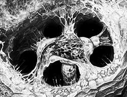
More detailed understanding of how physiologies develop and are maintained is attainable in zebrafish because of facile live-imaging, CRISPR-based genetics, gene-editing, and biophysical measurements. The integration of quantitative cell biology, developmental biology, and physiology will accelerate our progress toward a better understanding of how organs work and lead to new therapeutic strategies. Currently, we combine multi-scale imaging, genetics, and physical approaches to uncover the principles underlying the formation and function of tissue-scale pressure relief valves in the ear and the eye.
Current Projects
- How do developmental signals control the emergence of the ear’s relief valve?
- What mechanisms control pressure within the eye?
- What are the multi-scale mechanisms that establish and maintain organ form and function?
Selected Publications
Swinburne, I.A., Mosaliganti, K.R., Upadhyayula, S., Liu, T-L., Hildebrand, D.G.C., Tsai, T.Y.-C., Chen, A., Al-Obeidi, E., Fass, A.K., Malhotra, S., Engert, F., Lichtman, J.W., Kirchhausen, T., Betzig, E., and Megason, S.G. Lamellar projections in the endolymphatic sac act as a relief valve to regulate inner ear pressure. eLife 7:e37131 (2018).
Mosaliganti, K.R.*, Swinburne, I.A.*, Chan, C.U.*, Obholzer, N.D., Green, A.A., Tanksale, S., Mahadevan, L., and Megason, S.G. Size control of the inner ear via hydraulic feedback. eLife 8:e39596 (2019).
Liu, T.L.*, Upadhyayula, S.*, Milkie, D.E., Singh, V., Wang, K, Swinburne, I.A., Mosaliganti, K.R., Collins, Z.M., Hiscock, T.W., Shea, J., Kohrman, A.Q., Medwig, T.N., Damournet, D., Forster, R., Cunniff, B., Ruan, Y., Yashiro, H., Scholpp, S., Meyerowitz, E.M., Hockemeyer, D, Drubin, D.G., Martin, B.L., Matus, D.Q., Koyama, M., Megason, S.G., Kirchhausen, T., and Betzig, E. Observing the cell in its native state: imaging subcellular dynamics in multicellular organisms. Science 360, eaaq1392 (2018).
Xiong F., Ma W., Hiscock T.W., Mosaliganti K.R., Tentner A.R., Brakke K.A., Rannou N., Gelas A., Souhait L., Swinburne I.A., Obholzer N.D., and Megason S.G. Interplay of cell shape and division orientation promotes robust morphogenesis of developing epithelia. Cell 159(2): 415-27 (2014).
Xiong, F., Tentner, A.R., Huang, P., Gelas, A., Mosaliganti, K.R., Souhait, L., Rannou, N., Swinburne, I.A., Obholzer, N.D., Cowgill, P.D., Schier, A.F., and Megason, S.G. Specified neural progenitors sort to form sharp domains after noisy Shh signaling. Cell 153(3): 550-561 (2013).
Obholzer N*, Swinburne I.A.*, Scwab E, Nechiporuk A.V., Nicolson T, and Megason S.G. Rapid positional cloning of zebrafish mutations by linkage and homozygosity mapping using whole-genome sequencing. Development 139(22): 4280-90 (2012).
Swinburne, I.A., Miguez, D., Landgraf, D. and Silver, P.A. Intron length increases oscillatory periods of gene expression in animal cells. Genes & Development 22(17): 2342-6 (2008).
Swinburne, I.A., and Silver, P.A. Intron delays and transcriptional timing during development. Developmental Cell 14(3): 324-30 (2008).
Swinburne, I.A., Meyer, C.A., Liu, X.S., Silver, P.A., Brodsky, A.S. Genomic localization of RNA binding proteins reveals links between pre-mRNA processing and transcription. Genome Research 16(7): 912-21 (2006).
Brodsky, A.S., Meyer, C.A., Swinburne, I.A., Hall, G., Keenan, B.J., Liu, X.S., Fox, E.A., Silver, P.A. Genomic mapping of RNA polymerase II reveals sites of co-transcriptional regulation in human cells. Genome Biology 6(8): R64 (2005).
*Equal contribution
Last Updated 2020-08-17