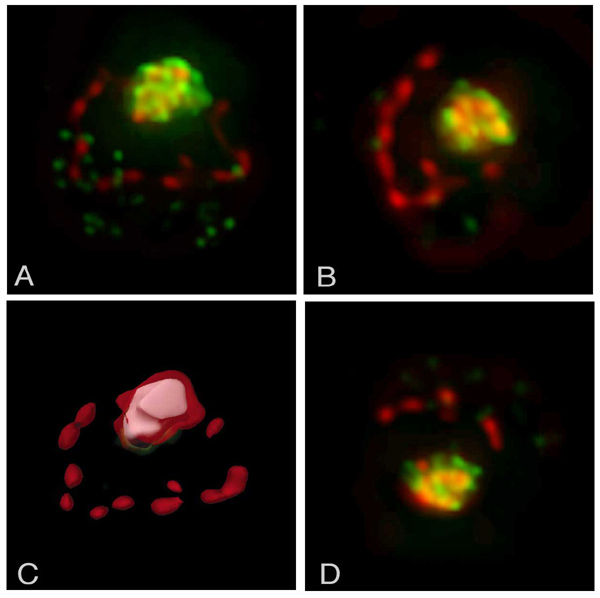
A YEATS-domain containing protein, Taf14p, is detected by polyclonal antibodies to Taf14p and anti-rabbit AlexaFluor 488 secondary antibody (red). Nuclei are stained with DAPI (green). This deconvolved image shows that Taf14p is efficiently localized to the nucleus in the absence of TFIIS. A, B, D: Individual yeast cells. C: Cell "A" with visible colocalization channel (white). These images were obtained using the DeltaVision Restoration microscope in the Biological Imaging Facility. Deconvolution was done using Huygens Pro software. Colocalization was done using Bitplane Imaris.
Photo courtesy of Professor Emerita Caroline Kane