 |
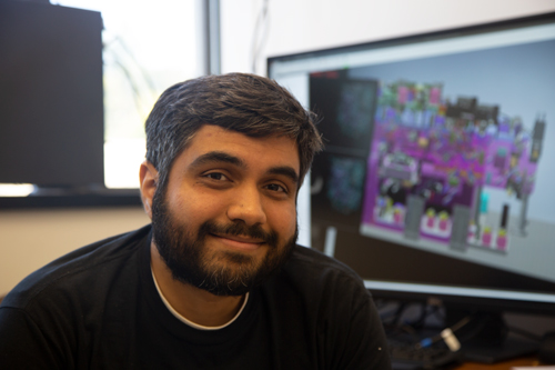
| MCB Assistant Professor in Residence and ABC Scientific Director Gokul Upadhyayula with the “Swiss Army Knife Microscope” schematic on the monitor behind him. Photo credit: Ann Trapaga |
We are excited to officially welcome new faculty member Srigokul ‘Gokul’ Upadhyayula to MCB! Upadyayula joins MCB as an Assistant Professor in Residence in the Division of Cell & Developmental Biology and is the first Scientific Director of the Advanced Bioimaging Center (ABC, formerly the Advanced Imaging Center) on campus.
Upadhyayula, who goes by Gokul, along with MCB faculty and ABC co-founders Eric Betzig, Xavier Darzacq, Doug Koshland, and Robert Tjian, aim to develop the ABC into a visualization, data processing, and analysis powerhouse. It will bring together scientists with broad specialties (e.g., applied mathematics, biology, computer science, instrumentation), give access to revolutionary imaging instruments and resources, and provide the support researchers need to visualize, analyze, and understand the data to extract scientifically meaningful insights.
Since the center was established in the spring of 2018, the facility in Barker Hall has had a 100Gbps network and mini-computing cluster (approx. 700 cores and 16 GPUs) installed and is working on hiring scientific staff. They have hired their first computational biologist postdoc, a Berkeley undergraduate to help set-up and support the lab, and are interviewing instrumentation scientists, who will help with building and maintaining the instruments. This summer two diligent high school students are also volunteering to help set-up the lab.
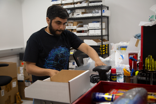 |
| Gokul preparing microscope components for assembly. Photo credit: Ann Trapaga |
Two cutting-edge adaptive optical multi-functional microscopes are also being built. Dubbed the "Swiss Army Knife Microscope," they combine multiple modes of imaging with integrated light paths. These microscopes are designed to seamlessly switch between modes of imaging to balance resolution, speed, invasiveness and imaging tradeoffs, which allows them to function more optimally with a more diverse set of biological specimens and conditions. This powerful technology allows biological processes and systems to be observed in a more natural environment in ways which have never been possible before. For example, specimens up to several millimeters in size can now be imaged over sessions lasting up to multiple days.
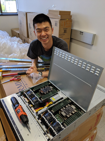 |
| UC Berkeley undergraduate, Aaron Sun, helps prepare and assemble a computing cluster. Photo credit: Gokul Upadhyayula |
The ABC’s goal is to have the first of the two microscopes operational this fall. They will then begin accepting project proposals with the eventual intent of establishing a global advisory committee that will review proposals from scientists interested in conducting experiments at the ABC on a bi-annual basis. These projects will help propel developments in emerging microscopy technologies as they will test the limits of the current instruments.
Gokul is keen to lead the ABC efforts to provide Berkeley researchers and eventually the broader scientific community access to cutting-edge microscopy and the resources and support needed to process the tera- and petabyte-scale projects, and develop robust computational workflows that allow researchers to extract meaningful and novel insights. He is "excited by the enthusiasm and the collaborative instinct of Berkeley faculty across disciplines" and is "very much looking forward to working with faculty across campus [who will] bring their complementing and synergistic expertise to projects at the ABC."
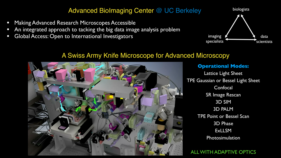 |
| An overview of the Swiss Army Knife Microscope abilities and ABC objectives. Slide courtesy: Gokul Upadhyayula |
| Watch these advanced imaging videos of live cells in action: | |
| Dynamics of cell division along the spine of a developing zebrafish embryo. The video is a ~2um volume projection where cell plasma membranes and golgi are shown in green, ER in red and mitochondria in blue. Courtesy Gokul Upadhyayula. | Immune cells on patrol in the endolymphatic space of a developing zebrafish embryo and imaged using the adaptive optical lattice light-sheet microscope (cells in green, and fluorescent sugars in magenta). Courtesy Gokul Upadhyayula. |