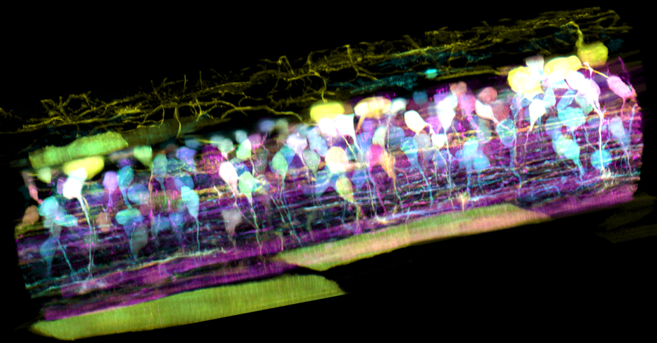
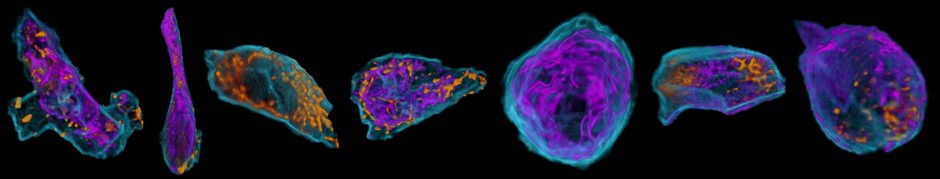
Image below: Organelles from the eye cell of a Zebrafish embryo.
The Advanced Imaging Center: Changing How We See
When Eric Betzig — winner of the 2014 Nobel Prize in chemistry for his work developing super-resolution fluorescence microscopy — joined Berkeley’s faculty last spring, he was drawn to the campus’s reputation as a global leader in imaging. He expressed hope that one day he could help create a state-of-theart imaging center that would attract scientists internationally and promote creative collaboration. That day has arrived.
Located on the third floor of Barker Hall, the Advanced Imaging Center (AIC) will enable Berkeley’s faculty to develop new microscope technologies that are in the pre-commercial stage, which will eventually become available to researchers worldwide. Betzig helped establish a similar center at the Janelia Research Campus in Virginia and will serve as an adviser to Berkeley’s facility. Xavier Darzacq — assistant professor of genetics, genomics, and development — and Doug Koshland — the Richard and Rhoda Goldman Distinguished Chair in the Biological Sciences and professor of genetics, genomics, and development — will help Betzig bring this vision to fruition.
“Very often, years pass between the first proof-of-concept of a new technology and its broad use and commercialization, assuming the technology ever survives that ‘valley of death,’” explains Koshland. The AIC initiative will help address challenges along the whole pipeline of research and development, from basic science to attracting the attention of commercial vendors and providing support for the development of new image-analysis tools. “All the fields of biology are undergoing a profound transformation in which imaging technologies can now bridge spatial scales ranging from single proteins to full organisms with temporal resolutions ranging from milliseconds to days,” Koshland says. “New measurements are changing how we see the different reactions happening in cells and how regulatory mechanisms operate. But although many technologies enable these measurements, they’re restricted to a handful of labs that are equipped to design and implement them.”
The initiative has certainly been strengthened by the presence of Betzig, who pioneered a profound transformation that opened the door to molecular resolved live cell imaging by inventing PALM (photo-activated localization microscopy) and developing lattice light sheet microscopy. He particularly hopes to link biological users, instrumentation operators and developers, and Bay Area data scientists to create a facility even more potent than at the Janelia center — especially by tackling the big-data analysis challenges presented by these cutting-edge microscopes.
The AIC expects to attract top biology researchers from around the world to collaborate across all scientific disciplines and could prove a useful tool for recruiting top-notch new faculty. Innovations in data analysis and mining tools will no doubt result from working with data scientists at Cal and throughout the Bay Area. “New technologies used by a few are bound to remain at the level of glorified, soon to be forgotten, proofs-of-concept,” says Darzacq. “The revolution can only be powered by the masses.”
The team concurs that the biggest hurdle facing the successful implementation of imaging technology is the quantity and sophistication of data analysis required to transform images into biological insight. “This hurdle can only be solved by the dynamic interactions of engineers, biologists, and computer scientists, both during and after image acquisition,” Betzig says. “The AIC will provide a unique cauldron for these dynamic interactions by attracting remarkable scientists in all these disciplines from Berkeley and beyond.”