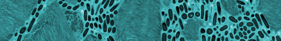
Image below: Retinal pigment granules are intermixed with rod and cone photoreceptor cells of the outer segment layer in a Zebrafish larva imaged by TEM.
The Electron Microscope Lab: A New Wave
The Advanced Imaging Center (AIC) isn’t the only imaging core and shared resource facility on campus. One of its collaborative partners is the Electron Microscope Laboratory (EML), which has been a fixture at Berkeley since the early 1960s and is now introducing a new wave of technology and capabilities for many types of sample preparation.
The EML’s primary mission is to serve as a teaching and training facility for the entire UC Berkeley community, says EML Director Danielle Jorgens (PhD 2013, Comparative Biochemistry). The lab offers project consultation, education and training for new users, equipment for trained users who anticipate long-term EM projects, and full-service microscopy. Faculty and students across campus — from chemistry to environmental studies to even the art departments — find their way to the north basement of Giannini Hall to take advantage of the state-of-the-art equipment. Whether it’s transmission EM, scanning EM, mounting specimens, or capturing images, the lab can tailor its training to the needs of each project.
Electron microscopy is undergoing a revolution due to techniques like cryo-electron microscopy and VolumeEM, which enable scientists to get higher-resolution results faster. Jorgens hopes to add powerful new microscopes in the future, such as Focused Ion Beam Scanning Electron Microscopy (FIB-SEM), capable of viewing samples at nanometer resolution across all three dimensions. The lab is also expanding its reach with Correlative Light and Electron Microscopy (CLEM), a technique that integrates fluorescence and electron microscopy datasets with ease and precision (shown in banner image on main page).
That’s where the AIC comes in — the two facilities are actively collaborating and generating data on CLEM projects. By closely partnering with scientists working on new imaging technologies at the AIC, the EML will help enable synergistic and fluid data collection between light and electron microscopy.
In addition to serving the campus community, Jorgens says, the EML works with private companies and other academic institutions worldwide. In this hybrid model as both a shared resource and a business, the lab is now offering a full-service option if researchers can’t devote their own time to an EM project.
The EML facility also teaches two MCB courses each semester, 481B and 481C. Marc Lim, a second-year bioengineering PhD student in Ting Xu’s lab, has attended both classes and trained at the EML to further his research on drug delivery to brain tumors. His work relies on fine spatial details such as collagen fibrils and blood vessel pores in the tumor micro-environment. “The EML staff gave me all the necessary tools to succeed,” he says, “from tissue preparation and treatment to procedural protocols.”
“At every level, we’re devoted to delivering excellent data,” Jorgens adds. “Whether it’s an undergraduate student just learning how to use the equipment or a PI who’s been doing EM for 20 years, our end goal is to help facilitate great science.”
For more information about the EML, visit their website.
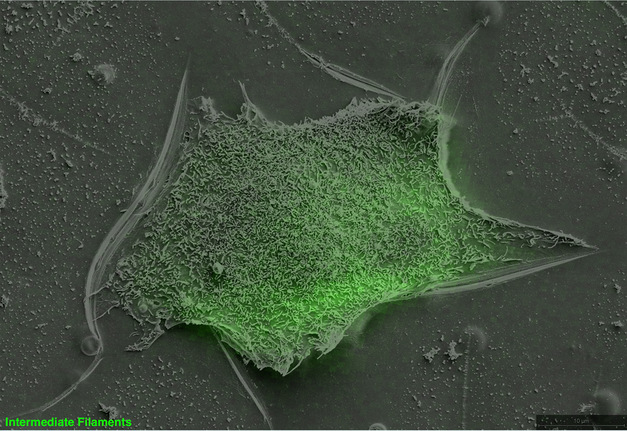
Image below: Correlative immunofluorescence and scanning electron microscopy image of a breast cancer cell.