MCB TRANSCRIPT
New Brain Microscopy Innovation Center:
Tracking Neural Activity in Living Animals
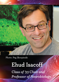
Researchers are closer than ever to finally understanding how our brains work, thanks to new tools that use light to manipulate and report on the activity of neurons and synapses in living animals. But commercial microscopes have not yet caught up with these advances, limiting their use among researchers who can't adapt or build their own microscopes. "Production of instruments and dissemination of know-how to enable use by the entire research community is a major need," says MCB Professor Ehud Isacoff.
Now, UC Berkeley offers a solution: a new center — the $5.6 million Brain Microscopy Innovation Center (BrainMIC) — to foster development of commercial microscopes that meet neurobiologists' needs. This partnership with leading microscope manufacturer ZEISS Microscopy celebrated its Grand Opening in May 2015, and UC Berkeley researchers are now adapting the most advanced microscopes currently available.
"Commercial microscopes are typically closed boxes whose warrantees can be invalidated by the mere thought of a screwdriver. ZEISS is providing us with the top-line new turnkey instruments as well as open access versions so we can play with them, and ZEISS will adopt and market our adaptations," Isacoff says. "This will benefit us, the company and the entire neuroscience community." The new center is part of the Helen Wills Neuroscience Institute, which Isacoff directs.
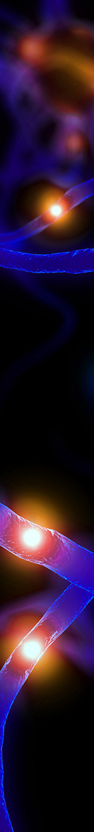
So far, BrainMIC has several top ZEISS microscopes. Features include superior image quality, images at greater depths, and faster scanning. A light sheet microscope, for example, illuminates all the neurons in a plane rather than just one point at a time. This allows researchers to scan an entire brain — like the 80,000-neuron brain of a larval zebrafish — faster than one can typically scan a single layer. "It's wonderful for imaging every neuron in the brain while the animal is alive and behaving," Isacoff says. "You can see all the neural activity nearly at once."
However, this light sheet microscope works only on transparent brains like those of fruit flies and larval zebrafish. The BrainMIC team wants to develop a similar microscope for the mouse brain, which is opaque. Currently, imaging an entire mouse brain requires a process of clearing that can only be done post-mortem. "We're working on three new strategies for microscopy for the mouse brain that penetrate deep into that opacity, effectively clearing the live brain," Isacoff says.
Starting in winter 2016, BrainMIC will offer a course on new brain microscope technologies that will be open to all. "The East Coast has similar courses in the summer but there has been nothing on the West Coast," Isacoff says. "This will create a new hub of microscope technology."
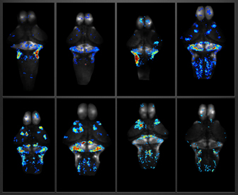
(activity increases from cool to hot colors).