MCB TRANSCRIPT
Brain Activity: Imaging Communication in Living Neurons
Assistant Professor Evan Miller gets the best of both worlds from his joint appointment in MCB and chemistry. “I love organic synthesis, and biology gives me a reason to do chemistry,” he says. “I’m building fluorescent sensors to study how neurons talk to each other.” This is a challenge because our brains are incredibly complex, with more than 80 billion neurons that make perhaps 10 trillion connections altogether.

Miller began making fluorescent molecules as a graduate student in chemistry and chemical biology at UC Berkeley. Then, as a post-doc, he developed a fluorescent dye that images neuronal activity, showing the tiny, fleeting changes in voltage across their cell membranes as they fire. “The dye lets you read light to detect activity — this is a new way of voltage sensing,” he says, explaining that the old ways were either fast or sensitive but not both at the same time.
In contrast to the conventional approach of sticking electrodes into neurons one by one, Miller’s dye enables tracing the activity of multiple neurons in a circuit simultaneously. Applications include imaging neurons in action in cell culture, tissue slices, and living animals. “I want to make imaging tools that will eventually let us watch movies of the whole brain,” he says.
Now, Miller is optimizing his voltage-sensing system. Besides giving a strong, responsive signal, his voltage-sensing dye had to strike a balance between water solubility (which makes it compatible with living cells) and lipid solubility (which keeps it mostly in the cell membrane). But the dye doesn’t extend all the way across the membrane, and Miller thinks increasing the dye’s reach could maximize its sensitivity.
He’s also expanding the dye palette, which would facilitate color-coding different neurons. More colors would also allow combining his technique with optogenetics, which uses light to turn neurons on and off. Currently, the two techniques use the same wavelength of light, making them incompatible. Combining them would let researchers use light both to stimulate neurons and to read the effects of that stimulation.
Another goal is developing fluorescent dyes that are triggered only in particular kinds of neurons. “You can target neurons in a specific developmental lineage, using a genetically encoded element to activate the dye,” he explains. This is already done with optogenetics, and he wants to piggyback his imaging system onto that.
Miller is glad to be back at UC Berkeley. “There are amazing people in many different fields,” he says. “It’s very exciting to be able to work with all these perspectives — insights from across disciplines drive what molecules we build and what biological questions we ask.”
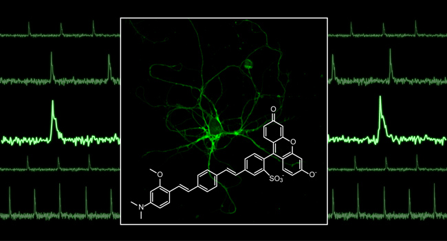
in real time in living neurons (green, center).