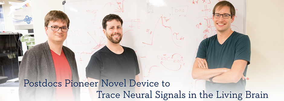
Below: Nicolas Pégard, Alan Mardinly and Ian Oldenburg (left to right) created the 3D-SHOT device, which uses laser light to stimulate neurons and trace the resulting signals throughout the brain (masthead image on Fall 2018 Transcript main page).
In the lab of MCB’s Hillel Adesnik, three postdocs with diverse academic backgrounds have made dramatic progress on instrumentation to enable the creation, manipulation, and editing of brain activity. Their work will help advance the fundamental research necessary to pursue treatments for dementia, Parkinson’s and other diseases of the brain.
Ian Oldenburg and Alan Mardinly knew each other at Harvard Medical School, where they both earned Ph.D.s in neuroscience, while Nicolas Pégard earned his Ph.D. in electrical engineering from Princeton. Their common interest in imaging neuronal activity was the impetus for collaborating on this project.
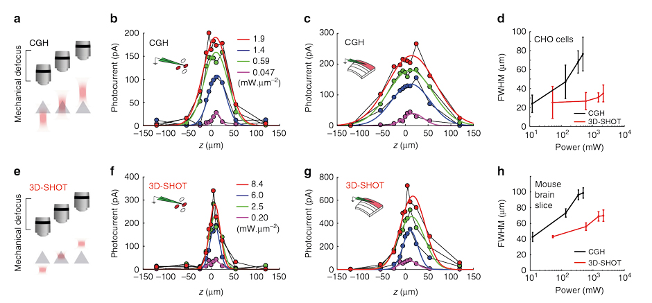
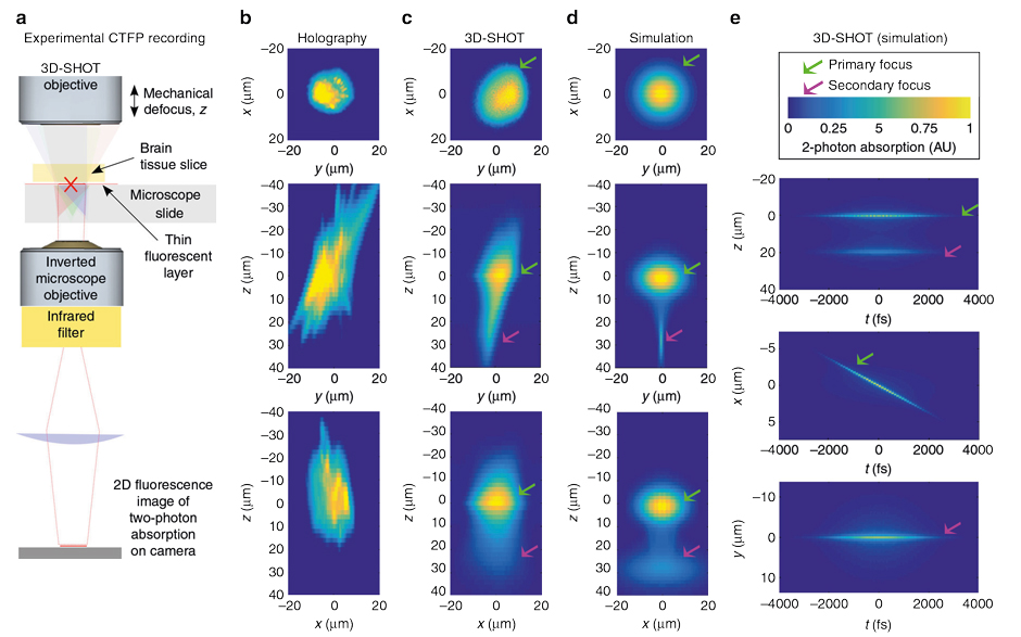
The team has created a technology they call 3D-SHOT with a laser that creates a miniaturized cloud of focused dots, flickering hundreds of times a second, to stimulate neuron ensembles on demand and trace the resulting signals propagating through the brain in interconnected networks involving thousands of neurons.
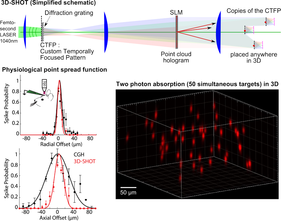
Says Pégard, “Reading and writing in an awake brain with light is extremely challenging and cannot be done efficiently with off-the-shelf microscopes. As a team, we designed the optical system and the software exclusively for one task: communicating with neurons at high speeds. Our goal was to create the most reliable toolbox for neuroscientists to build upon. With 3D-SHOT, neuroscientists can read and write in the brain, and can start cracking the neural code with a reliable all-optical interface to the brain.”
“Using 3D-SHOT and new light-sensitive proteins called opsins, we can successfully create custom patterns of brain activity, but we are just at the beginning of learning about how brains work. Therapies for brain diseases based on these approaches might be decades away. The immediate application of these approaches will be in basic neuroscience research, where it will allow scientists to ask new types of questions about how activity in the brain encodes actions and sensation,” adds Mardinly.
In 2017, the three researchers proposed a conference they called “Sculpted Light in the Brain.” They started with a $2,500 grant from QB3, invited speakers and provided free registration. As Oldenburg explains, “We decided to start the sculpted light meetings because we really wanted a venue to talk to all the other people in the world who are thinking about these types of technological problems. Solving these problems requires optical engineers, molecular biologists and basic neuroscientists all working on the same goal."
Three hundred people registered, all the invited speakers attended, and the three organizers raised a total of $20,000 from foundations and other sponsors to cover travel and other expenses. Adds Oldenburg, "We were overwhelmed by how many people shared this idea and all the positive response.”
The team has created a non-profit organization to continue organizing the conferences. The next one will be in London in 2019.
More information is available at sculptedlight.org