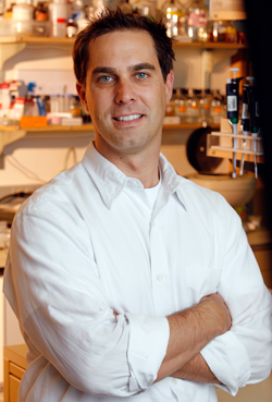
Andrew Dillin

Howard Hughes Medical Institute Investigator and Thomas and Stacey Siebel Distinguished Chair in Stem Cell Biology and Professor of Immunology and Molecular Medicine
Lab Homepage: https://www.dillinlab-berkeley.org/Research Interests
As an organism ages, its proteins face an increasing severity in the challenges they receive from extrinsic and intrinsic environmental perturbation. Chaperones become dysregulated, while the degradation machineries stop working properly. The protein accumulates damage and starts to misfold. At this point, the cell needs to mount a response to restore its homeostasis; however, the stress response machinery that it typically relies upon when faced with such challenges has lost its capacity to function.
This breakdown, however, does not lead to complete disorder. As the organism ages, it exhibits a degree of correlated, recognizable, and predictable changes to its physiology over time. These changes can occur synchronously across multiple tissues and organs. The phenotypic changes of aging occur in a type of concert, rather than in isolation, suggesting the residual participation of the endocrine system in the onset of age-related phenotypes. The demise of the cell thus most often occurs within the context of the simultaneous demise of the whole organism.
Our lab focuses on the questions of why an aging organism begins to lose control over the integrity of its proteome, and how this loss is communicated across its various tissues. To accomplish this, we have taken the approach of breaking down a cell into its small and canonically-autonomous parts – its suborganelles and subcompartments – such that we can take a larger step back to ask how those smaller portions can communicate both with each other and with the organism as a whole. Our approaches have required us to diversify the systems in which we ask questions: we work on model systems ranging from stem cells and nematodes to mice. We have developed and applied techniques that allow us to manipulate signaling pathways or proteins within a single tissue, cell, or an organelle within a single cell so that we can observe how that small perturbation might reverberate and effect the physiology of the whole of the organism. Our work is fundamentally grounded in the endocrinology and genetics of aging, and our larger goal is to apply our findings towards uncovering new therapeutic strategies for the treatment of age-related pathologies.
Current Projects
The Communication of Mitochondrial Proteotoxic Stress
Mitochondrial dysfunction has been reported as a cause or as a consequence of nearly every single age-onset human disease. Recent work within our lab suggests that mitochondria potentially communicate or actively signal to regulate one another's activity between tissues. We have found that a tissue-specific mitochondrial stress can be sensed and transmitted to distal cells, invoking a cell non-autonomous mitochondrial stress response that extends life span. We are now working toward understanding the source and nature of this signal and the functional and morphological consequences of its signaling.
The Communication of Endoplasmic Reticulum Stress
The endoplasmic reticulum (ER) is responsible for the folding and maturation of up to as many as 13 million proteins per minute. Challenges to the ER folding environment can have a multitude of consequences on an organism’s viability: defects in ER function are strongly associated with a large number of metabolic and age-onset disorders. We wish to understand how ER stress is signaled and perceived throughout the organism as it ages, and the extent to which such signaling is conserved in vertebrates.
The Function of Cytoplasmic Heat Shock Response in Aging
We have an additional team within the lab working on stress responses of the cytoplasm. The idea behind this work is to understand how the cytoplasmic stress responses might affect proteome maintenance and the rate of aging of the entire organism. The master regulator of the cytoplasmic stress response is the transcription factor HSF-1. We have isolated a hypermorphic variant of HSF-1 that can dramatically extend the life span of the nematode and increase thermotolerance. Paradoxically, it does not result in a canonical upregulation of the downstream chaperone network typically ascribed to the achievement of these phenotypes. Recently, we have created a mouse model in which HSF-1 is constitutively activated in the CNS, and we are actively working to understand HSF-1’s effects on proteotoxic stress in this background. Importantly, this body of work may reveal novel aspects of heat shock regulation, conserved from worms to mice, that function independently of chaperone induction.
Stem Cells, Nutrient Availability and the Aging Process
The extreme pressure that evolution places on reproductive success has long suggested that resources will be allocated differently across cell types: for example, to ensure that the germline retains a pristine health that assures the survival of the organism’s progeny, resources required for a heightened maintenance of cellular defenses must be upregulated. Elimination of reproduction would conversely signal the upregulation of defense mechanisms in somatic tissue, and dietary restriction is likewise thought to re-appropriate resources from reproduction towards somatic cell defenses. We thus hypothesize that germline cells (such as human embryonic stem cells) or nutrient-deprived cells might exhibit a heightened capacity to ensure proteostasis and thereby avoid protein damage. Accordingly, we have invested a considerable amount of effort in understanding how the proteome of stem and germ cells can remain pristine. Our work has focused on the functional consequences of the upregulation of the proteasome found in these cells.
In parallel projects, we have found that genetic manipulation of specific receptors in olfactory neurons in both mice and worms is sufficient to increase health span and longevity, and are actively working to uncover the genetic signaling pathway underlying this response. Our lab has also discovered a highly conserved transcription factor, PHA-4, required for dietary restriction in C. elegans. We believe that this protein forms a core-signaling pathway that responds to and integrates an organism's response to reduced caloric intake. We are thus working toward understanding the molecular mechanism by which this pathway perceives and interprets the environmental signals that ultimately result in increased longevity.
Understanding the Effects of Aging on the Translatome
We are actively working on applying emergent technologies to manipulate and analyze the workings of the translational apparatus throughout the whole organism and within specific tissues. Significantly, this includes the establishment of ribosome profiling methodologies in worms and in stem cells. These techniques will allow us to dissect translation at a nucleotide resolution as it occurs, and will provide fundamental insights into the effects of aging on the rate, pausing, and non-coding start sites of translation. Significantly, this system will allow us the capacity to both temporally and spatially disect apart the contributions of stress responses to changes in the translatome.
Selected Publications
Peer Reviewed Journal Articles:
Vilchez D, Boyer L, Morantte I, Lutz, M, Merkwirth C, Joyce D, Spencer B, Page L, Masliah E, Berggren WT, Gage FH, and Dillin A. Regulation of FOXO4 and PSMD11/rpn-6 determines proteasome activity and human stem cell function. Nature. 2012: 489(7415):304-8. PMCID: 22972301.
Vilchez D, Morantte I, Liu Z, Douglas PM, Merkwirth C, Rodrigues, APC, Manning G, and Dillin A. RPN-6/PSMD11 is a determinant of C. elegans longevity and proteasomal activity. Nature. 2012:389(7415):263-268. PMCID: 22922647.
Mair W, Morantte I, Rodrigues AP, Manning G, Montminy M, Shaw RJ, Dillin A. Lifespan extension induced by AMPK and calcineurin is mediated by CRTC-1 and CREB. Nature. 2011;470(7334):404-8. PMCID: 3098900.
Durieux J, Wolff S, Dillin A. The cell-non-autonomous nature of electron transport chain-mediated longevity. Cell. 2011;144(1):79-91. PMCID: 3062502.
Cohen E, Du D, Joyce D, Kapernick EA, Volovik Y, Kelly JW, Dillin A. Temporal requirements of insulin/IGF-1 signaling for proteotoxicity protection. Aging Cell. 2010;9(2):126-34. PMCID: 3026833.
Mair W, Panowski SH, Shaw RJ, Dillin A. Optimizing dietary restriction for genetic epistasis analysis and gene discovery in C. elegans. PLoS One. 2009;4(2):e4535. PMCID: 2643252.
Cohen E, Paulsson JF, Blinder P, Burstyn-Cohen T, Du D, Estepa G, Adame A, Pham HM, Holzenberger M, Kelly JW, Masliah E, Dillin A. Reduced IGF-1 signaling delays age-associated proteotoxicity in mice. Cell. 2009;139(6):1157-69.
Carrano AC, Liu Z, Dillin A*, Hunter T. A conserved ubiquitination pathway determines longevity in response to diet restriction. Nature. 2009;460(7253):396-9. PMCID: 2746748. * Corresponding author
Panowski SH, Wolff S, Aguilaniu H, Durieux J, Dillin A. PHA-4/Foxa mediates diet-restriction-induced longevity of C. elegans. Nature. 2007;447(7144):550-5.
Dong MQ, Venable JD, Au N, Xu T, Park SK, Cociorva D, Johnson JR, Dillin A, Yates JR, 3rd. Quantitative mass spectrometry identifies insulin signaling targets in C. elegans. Science. 2007;317(5838):660-3.
Cohen E, Bieschke J, Perciavalle RM, Kelly JW, Dillin A. Opposing activities protect against age-onset proteotoxicity. Science. 2006;313(5793):1604-10.
Wolff S, Ma H, Burch D, Maciel GA, Hunter T, Dillin A. SMK-1, an essential regulator of DAF-16-mediated longevity. Cell. 2006;124(5):1039-53.
Raices M, Maruyama H, Dillin A, Karlseder J. Uncoupling of longevity and telomere length in C. elegans. PLoS Genet. 2005;1(3):e30. PMCID: 1200426.
Hansen M, Hsu AL, Dillin A, Kenyon C. New genes tied to endocrine, metabolic, and dietary regulation of lifespan from a Caenorhabditis elegans genomic RNAi screen. PLoS Genet. 2005;1(1):119-28. PMCID: 1183531.
Venable JD, Dong MQ, Wohlschlegel J, Dillin A, Yates JR. Automated approach for quantitative analysis of complex peptide mixtures from tandem mass spectra. Nature Methods. 2004;1(1):39-45.
Dillin A, Hsu AL, Arantes-Oliveira N, Lehrer-Graiwer J, Hsin H, Fraser AG, Kamath RS, Ahringer J, Kenyon C. Rates of behavior and aging specified by mitochondrial function during development. Science. 2002;298(5602):2398-401.
Dillin A, Crawford DK, Kenyon C. Timing requirements for insulin/IGF-1 signaling in C. elegans. Science. 2002;298(5594):830-4.
Arantes-Oliveira N, Apfeld J, Dillin A, Kenyon C. Regulation of life-span by germ-line stem cells in Caenorhabditis elegans. Science. 2002;295(5554):502-5.
Dillin A, Rine J. Roles for ORC in M phase and S phase. Science. 1998;279(5357):1733-7.
Dillin A, Rine J. Separable functions of ORC5 in replication initiation and silencing in Saccharomyces cerevisiae. Genetics. 1997;147(3):1053-62. PMCID: 1208233.
McCracken AA, Karpichev IV, Ernaga JE, Werner ED, Dillin AG, Courchesne WE. Yeast mutants deficient in ER-associated degradation of the Z variant of alpha-1-protease inhibitor. Genetics. 1996;144(4):1355-62. PMCID: 1207689.
Loo S, Laurenson P, Foss M, Dillin A, Rine J. Roles of ABF1, NPL3, and YCL54 in silencing in Saccharomyces cerevisiae. Genetics. 1995;141(3):889-902. PMCID: 1206852.
Fox CA, Loo S, Dillin A, Rine J. The origin recognition complex has essential functions in transcriptional silencing and chromosomal replication. Genes & Development. 1995;9(8):911-24.
Reviews, Perspectives, and Books:
Wilkinson DS, Taylor RC, Dillin A. Analysis of aging in Caenorhabditis elegans. Methods Cell Biol. 2012;107:353-81.
Taylor RC, Dillin A. Aging as an event of proteostasis collapse. Cold Spring Harb Perspect Biol. 2011;3(5).
Dillin A, Cohen E. Ageing and protein aggregation-mediated disorders: from invertebrates to mammals. Philos Trans R Soc Lond B Biol Sci. 2011;366(1561):94-8. PMCID: 3001306.
Douglas PM, Dillin A. Protein homeostasis and aging in neurodegeneration. J Cell Biol. 2010;190(5):719-29. PMCID: 2935559.
Dillin A. Andy Dillin: Using aging research to probe biology. Interview by Caitlin Sedwick. J Cell Biol. 2010;189(4):616-7. PMCID: 2872906.
Powers ET, Morimoto RI, Dillin A, Kelly JW, Balch WE. Biological and chemical approaches to diseases of proteostasis deficiency. Annu Rev Biochem. 2009;78:959-91.
Panowski SH, Dillin A. Signals of youth: endocrine regulation of aging in Caenorhabditis elegans. Trends Endocrinol Metab. 2009;20(6):259-64.
Mair W, Dillin A. Aging and survival: the genetics of life span extension by dietary restriction. Annu Rev Biochem. 2008;77:727-54.
Cohen E, Dillin A. The insulin paradox: aging, proteotoxicity and neurodegeneration. Nature reviews Neuroscience. 2008;9(10):759-67. PMCID: 2692886.
Balch WE, Morimoto RI, Dillin A, Kelly JW. Adapting proteostasis for disease intervention. Science. 2008;319(5865):916-9.
Durieux J, Dillin A. Mitochondria and aging: dilution is the solution. Cell Metab. 2007;6(6):427-9.
Wolff S, Dillin A. The trifecta of aging in Caenorhabditis elegans. Experimental gerontology. 2006;41(10):894-903.
Aguilaniu H, Durieux J, Dillin A. Metabolism, ubiquinone synthesis, and longevity. Genes & development. 2005;19(20):2399-406.
Dillin A, Rine J. On the origin of a silencer. Trends Biochem Sci. 1995;20(6):231-5.Enter a selected list of publications.
Photo courtesy of HHMI, credit Denis Poroy.
Last Updated 2012-11-16