

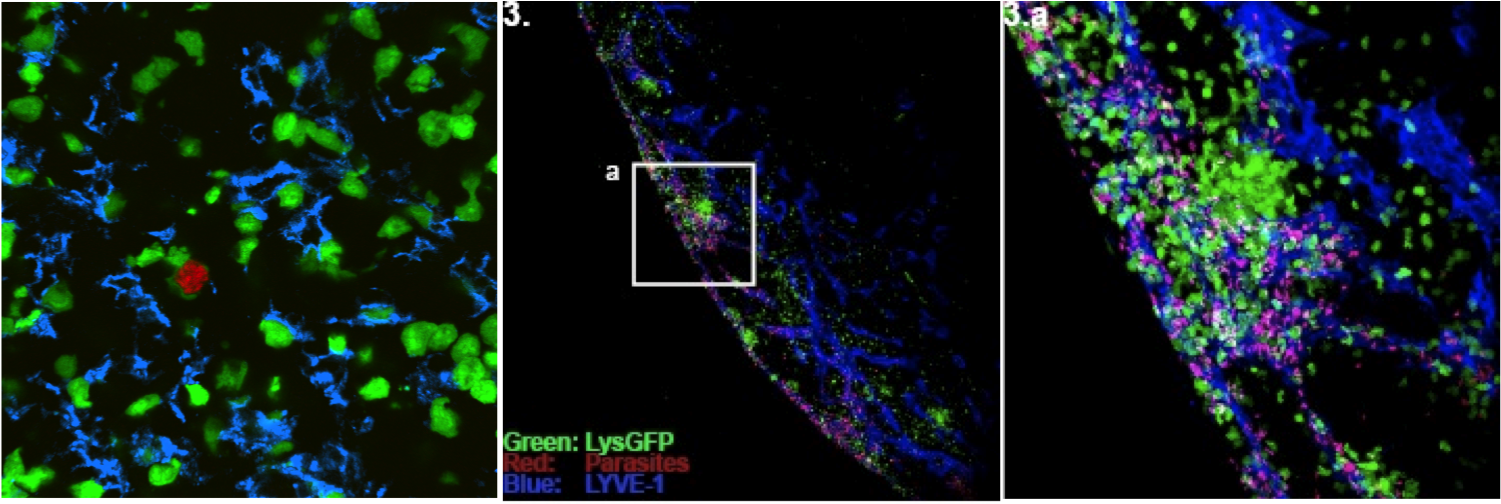
Lymphocytes exist within highly organized cellular environments. These tissue environments serve to segregate cells into different compartments, provide extracellular matrix for lymphocytes to crawl on, and help to present guidance and signaling molecules. Studies of lymphocytes removed from their normal environments provide an essential foundation for understanding the cellular responses to different stimuli. A full understanding of lymphocyte biology, however, will require integrating the information obtained from studies of isolated lymphocytes with studies of how the cells behave in their normal tissue environments.
In the past few years, the application of multiphoton imaging to analysis of lymphoid tissue has made it possible for the first time to perform real-time analysis of cells within their normal tissue environments.
Why use 2-photon microscopy?
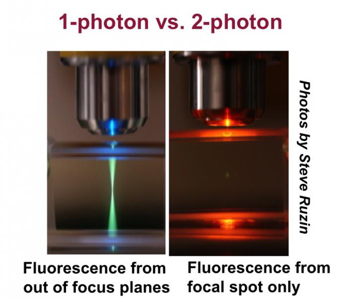
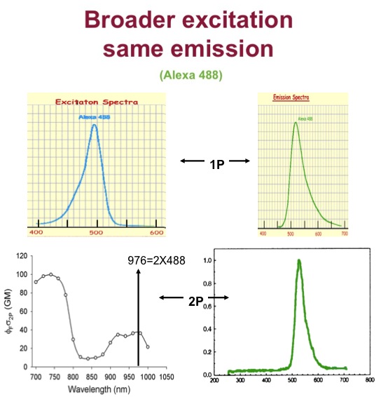
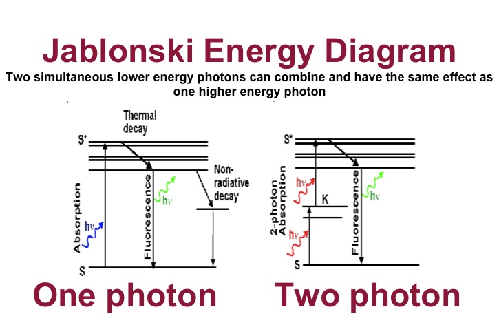
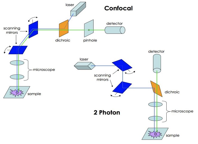
For questions that require imaging live cells for extended time periods deep within tissues, two-photon microscopy is the current method of choice. Like confocal microscopy, two-photon microscopy uses a laser to excite a fluorescent tag within a sample and detectors to measure the emitted light. However, unlike the lasers used for confocal microscopy, which provide single-photon excitation, the lasers used in two-photon microscopy excite by using near simultaneous absorption of two long wavelength (∼800 nm) photons. This leads to several distinct benefits. The long wavelengths used for two-photon microscopy are less damaging and penetrate more deeply into tissues than those used in confocal microscopy. In addition, the requirement for near simultaneous absorption of two photons means that excitation is only achieved near the focal plane where the laser light is most concentrated. This has two distinct advantages. One is that there is little tissue damage to the regions above and below the focal plane that are not being imaged. In addition, there is no out-of-focus light that could make the image blurry and difficult to interpret.
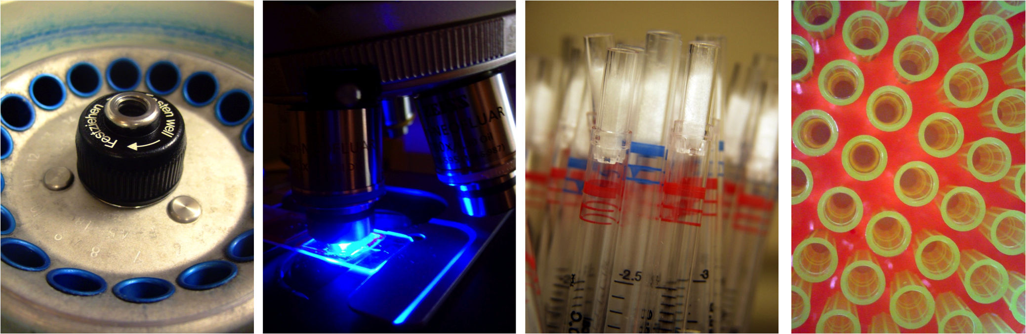
The 2-photon microscope
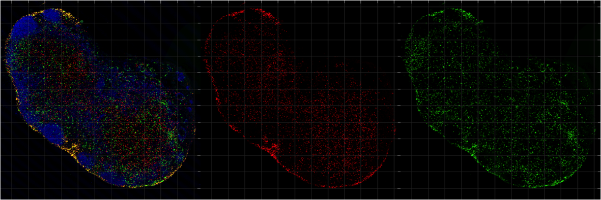
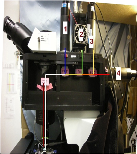
Two photon microscopes can look deep into fluorescent samples, typically 5-20 times deeper than other types of fluorescent microscopes. Our 2-P microscope can image four simultaneous color channels at 30 frames/second. It is well automated with computer control of the focus, stage motion and timing. A typical movie can show the cell movements in a 170 x 140 x 200 μm volume during 30 minutes.
Infra-red light is scanned across The main dichroic separates Four cameras detect four colors the sample 30 times per second the excitation from the emitted light simultaneously
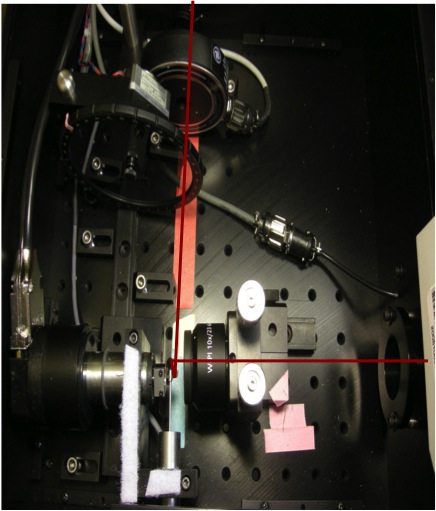
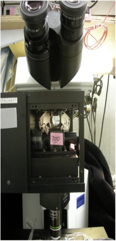
The simple light path of the 2-photon microscope makes construction easier:
Aligning the laser is easy and safe!


Please note that this photo was generated with Photoshop. Nobody was harmed during laser alignment. ;)
Please see the following helpful links:
Paul Herzmark's introduction to microscopy
Microscope questions
How to find difficult samples