Laboratory  of  John G. Forte

Department of Cell & Molecular Biology,  University of California, Berkeley


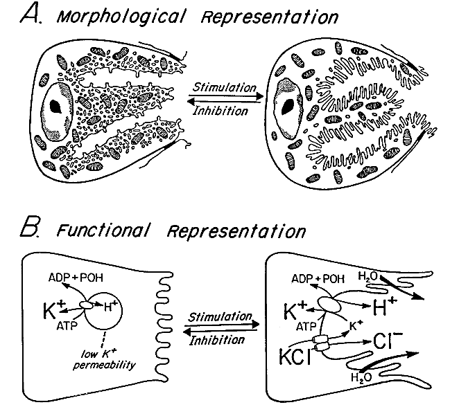
Morphological and functional transformation of the      
gastric parietal cell between resting and secreting states      

In the parietal cell's non-secreting state, the proton pumps (H,K-ATPase) are mainly in a complex intracellular membrane compartment consisting of many small vesicles having spherical, elongated or flattened shapes and commonly called "tubulovesicles."  These membranes lack a potassium (K+) permeability, blocking activity of the H,K-ATPase, which requires K+ at its extracytoplasmic face to transport in exchange for a proton (H+). (See more about tubulovesicles.)  The apical membrane, which does have channels for K+ and Cl-, forms multiply branched involutions, called "canaliculi" (greatly simplified here), emanating from a small apical pore, extending deep into the cell, and studded with microvilli.  A very high concentration of filamentous actin is closesly associated with the apical membrane, both in microvillar cores and in a subapical web.  (See more about this unusual apical membrane.)


Upon stimulation, many of the tubulovesicles fuse with the canalicular membrane, moving the proton pumps to a position in which they can actively exchange H+ for K+. This stimulation involves a massive movement of membrane to the canaliculi, distending them, elongating their microvilli, and greatly expanding the apical surface area.  The extent of membrane translocation per unit time is more extreme here than in any other cell in the human body, making the parietal cell an interesting model for examination of the processes of membrane fusion and endocytosis.
Protons transported to the canalicular lumen are joined by electrically balancing Cl- ions* and osmotically drawn water, all together constituting isotonic hydrochloric acid (~0.15N HCl) which drains through the lumen of each gastric gland into the cavity of the stomach.
 
 
* Cl- secretion involves the parietal cell's mechanism for dealing with the base (OH-) left behind by H+ secretion.  The protons being secreted come from ionization of water (H2OOH- + H+) and a growing imbalance of OH- over H+ would raise cytoplasmic pH to a point that would be lethal to the cell, so the excess base must be removed or neutralized.  Actively secreting parietal cells take CO2 from the blood (and from their own metabolism) and use the carbonic anhydrase enzyme to react it quickly with water, forming H+ and HCO3-.  The former replaces the secreted H+, maintaining a cytoplasmic H+/OH- balance, while the latter, a safer weak base (bicarbonate), rapidly leaves the cell across the basolateral membrane (to the blood), in exchange for Cl-, via anion exchangers (also called chloride/bicarbonate exchangers).  This entry of Cl-, driven above its equilibrium concentration in the cytoplasm by exit of HCO3-, provides the driving force for Cl- to cross the apical membrane as the anionic component of hydrochloric acid.

The process shifts the excess base from parietal cells to the blood, which is better able to handle the excess.  A much larger amount of bicarbonate already in the blood (part of the CO2/HCO3- buffering system), the buffering capacity of blood proteins, and automatic adjustments in breathing rate (to modify loss of CO2) keep the rise in blood pH very small when parietal cells are maximally secreting acid into the stomach and bicarbonate into the bloodstream.

Acid and base are only temporarily separated.  When acidic stomach contents empty into the duodenum, bicarbonate, secreted as sodium bicarbonate by the pancreas and duodenal lining, neutralizes HCl to form carbon dioxide, salt, and water:
NaHCO3 + HCl CO2 + NaCl + H2O