Laboratory of John G. ForteDepartment of Cell & Molecular Biology, University of California, Berkeley |
"Tubulovesicles" (or "tubulocisternae") — |
|
|
|
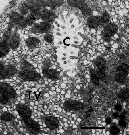
| TEM of resting parietal cell from piglet stomach, showing canaliculus (C) surrounded by numerous tubulovesicles (TV) and mitochondria. |
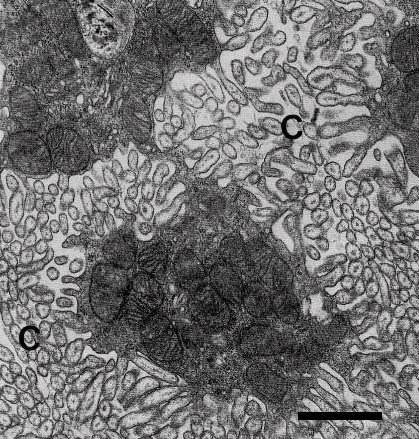
TEM of maximally stimulated parietal cell (piglet), showing greatly enlarged canaliculi with elongated microvilli. Remaining space has densely packed mitochondria and very few tubulovesicles. |
From: |
|
| HANDBOOK OF PHYSIOLOGY — THE GASTROINTESTINAL SYSTEM III, 1989 | |
Chapter 11: Cell biology of hydrochloric acid secretion |
| John G. Forte and Andrew Soll |
| Recently, high-pressure freezing was used to obtain better preservation of fine subcellular morphology. When serial thin sections were then used to reconstruct 3-dimensional shapes of the small membranous structures long called "tubulovesicles," the vesicles were seen to occur mostly as stacks of flattened cisternae. The term, "tubulocisternae," was coined to designate these structures, but long familiarity favors continued reference to "tubulovesicles." These 3-dimensional reconstructions add support for the view that the membranes containing most of the |
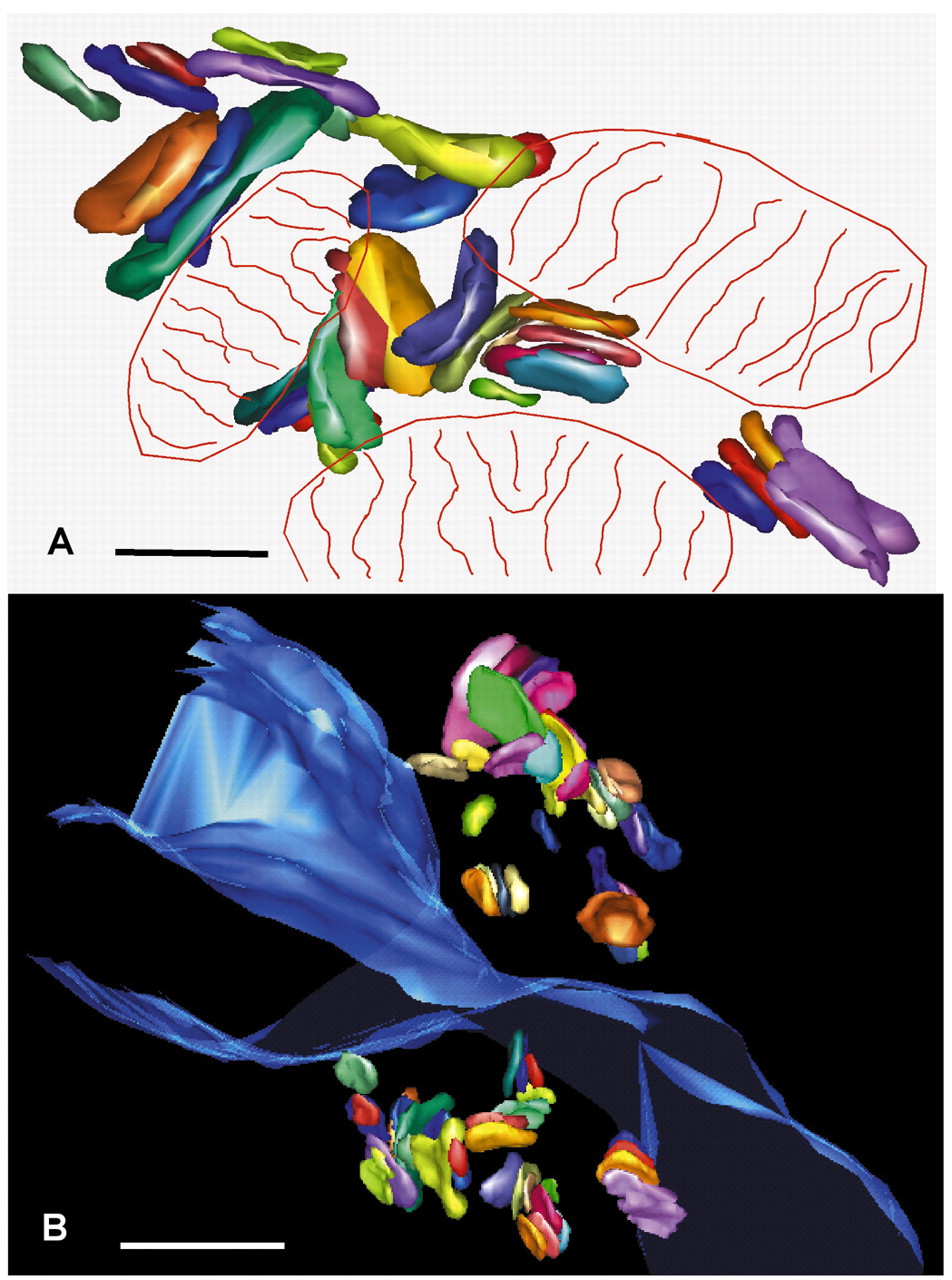 |
From Duman, et al., J Cell Sci. 2002: A. 3-D reconstruction of "tubulocisternae" in rabbit parietal cells, from serial thin sections after high-pressure freezing and freeze-substitution. Each membrane-bounded structure determined to have no membrane continuity with a neighboring structure was given a distinct color. Crude outlines of mitochondria are included. B. Lower-resolution 3-D reconstruction of a region of tubulocisternae surrounding part of a canaliculus (cut-away blue sheet structure). (Click for larger image in new window.) |