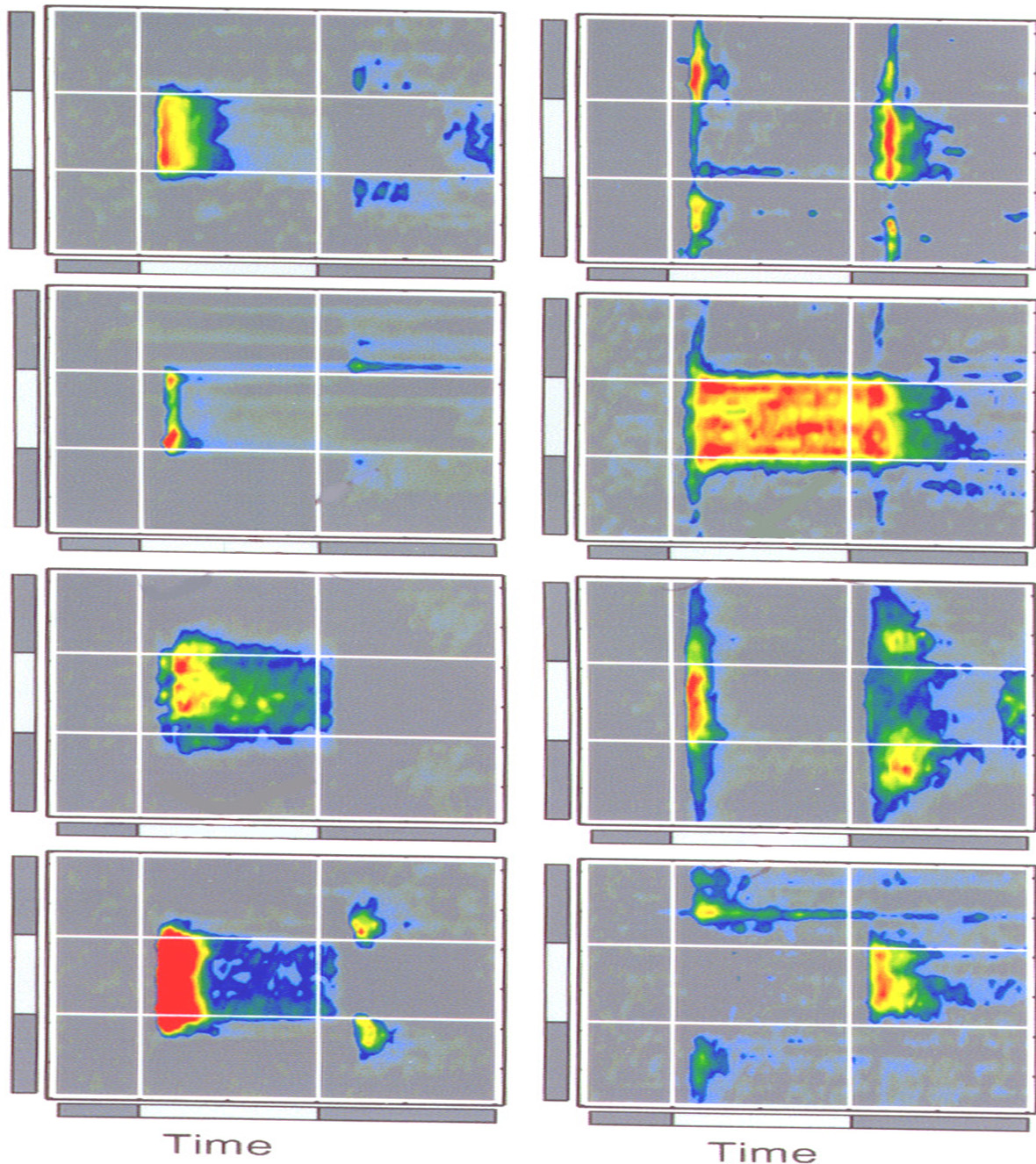

 Here are examples of 4 ON cells. Excitation on the left, inhibition on the right. Vertical white lines indicate stimulus ON and OFF. One second duration. Horizontal lines indicate stimulus width: 600 microns.

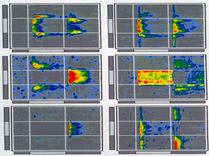
Here are similar rasters for 3 OFF cells. Note that, except for the LED, excitation and inhibition are complementary in both space and time.
Here are seven different representations of a face after processing by seven different retina filters. Each representation is shown in a different color. When the representations are merged, later in the movie, each color falls at a different region of the face, showing that each filter extracts a different region of the original scene. See how each of these representation corresponds to a different space-time domain in the frequency realm.
In concept, 30 electrodes record from a linear array of 30 ganglion cells of the say type, each spaced at 60 um. This recording generates a space-time representation of the original 600 um stimulus. It is shown as a color-coded raster in the final display. See the rasters in this view!!

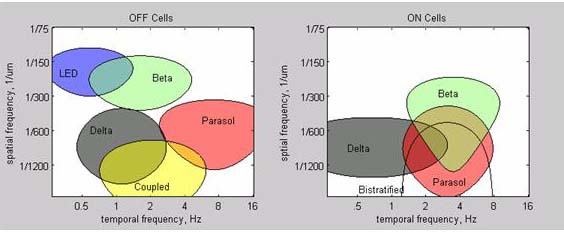
We approximated the spatial and temporal bandpass characteristics for each of the ganglion cell types measured by the raster technique. Here are the regions of activity for each cell type. The OFF cells seem to form discrete and separate regions that together span the full space-time domain. The ON cells are more problematical. Some of the specialized cells like the DS and Bistratified cells do not separate so neatly.
Cartoon of the retina showing layering of neurons
Feedback and Crossover inhibition
Multiple Representations of the Visual Scene
Regions of space/time frequency
Space-time rasters for ON and OFF cells
Patching a neuron in a retinal slice
Targeting Retinal Neuron Subregions with Arficial Rhodopsins