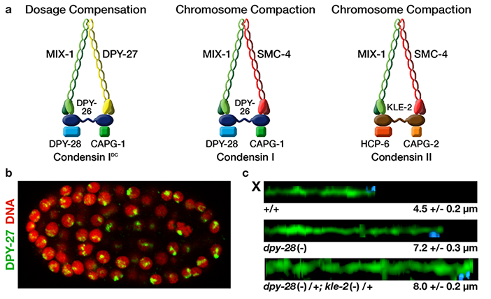
Figure 3: Biochemically distinct
condensin complexes with
interchangeable subunits control
chromosome structure
throughout C. elegans
development. a, Each condensin
complex contains a pair of SMC
proteins (Structural Maintenance of
Chromosomes) and three non-SMC
subunits. SMC proteins have
nucleotide binding domains at
their globular N- and C-termini,
which are linked by two long coiled
coil domains separated by a hinge
region. The dosage compensation
condensin complex (condensin IDC)
specifically changes the molecular
topology of interphase X
chromosomes in XX embryos to
repress gene expression by half. b,
An XX embryo co-stained with a
DNA dye and the DCC-specific
subunit DPY-27 shows X-specific
localization of the DCC. Condensin
I and II perform independent roles
in the structure and segregation of
mitotic and meiotic chromosomes,
with condensin II being the primary
regulator of chromosome
compaction and resolution (see
also h). Both condensin I and II
affect the number and distribution
of double strand breaks in meiosis,
and thereby the number and
position of crossovers between
homologous chromosomes, by
controlling the higher-order
chromosome structure. c,
Confocal images show that
mutations disrupting condensin I
and II subunits cause an additive
extension of chromosome axes.
Meiotic X-chromosome axes from
paired and synapsed homologs in
pachytene nuclei of wild-type and
condensin-mutant gonads were
stained with antibodies to the axial
protein HTP-3, traced in 3
dimensions, and computationally
straightened. Axial length is shown.
Condensin II co-localizes with
centromere proteins and mediates
faithful chromosome segregation.
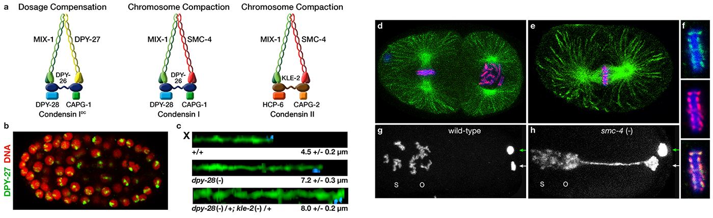
d, Two-celled, wild-type embryo
stained with DNA dye (blue) and
antibodies to tubulin (green) and
the centromere-specific histone
HCP-3 (red). The cell on the left is
in metaphase of mitosis. e,
One-celled, wild-type embryo in
metaphase stained with DNA dye
(blue) and antibodies to tubulin
(green) and the condensin II
subunit SMC-4 (red). f, Enlargement
of mitotic chromosomes in
metaphase co-stained with DNA
dye (blue) and antibodies to SMC-4
(green) and HCP-3 (red). The
merged image (bottom) shows the
colocalization of centromere
proteins and condensin II on the
holocentric nematode
chromosomes. DNA-stained
wild-type (g) and smc-4(-) (h)
embryos after segregation of
homologs (polar body, green
arrow) and sister chromatids (polar
body, white arrow) and migration
and fusion of the sperm (s) and
oocyte (o) pronuclei. In smc-4(-)
embryos the oocyte pronucleus
remains connected to the sister
chromatids in the polar body,
demonstrating improper
chromosome segregation in
meiosis. The dosage
compensation subunit MIX-1 is
shared with condensin II and
behaves similarly to SMC-4 in
mitosis.