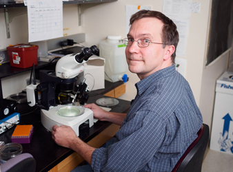
Craig T. Miller

Associate Professor of Genetics, Genomics, Evolution, and Development*
*Judy Chandler Webb Endowed Chair,
Research Interests
We study how pattern forms during development and changes during evolution. We focus on the vertebrate head skeleton, using a genetic approach in the threespine stickleback fish, a species complex that has repeatedly evolved head skeletal adaptations. We seek to understand the genetic basis of craniofacial and dental pattern and how alterations to these genes result in evolved differences in morphology.
Current Projects
The threespine stickleback fish (Gasterosteus aculeatus) has emerged as a powerful model system for studying the genetic basis of organismal diversity. Threespine sticklebacks have undergone one of the most recent and remarkable adaptive radiations on earth. Ancestral ocean-dwelling sticklebacks repeatedly colonized and rapidly adapted to thousands of freshwater lakes and streams that formed by melting of glacial ice within the past 15,000 years. Ancestral and derived forms can be crossed in the lab, enabling forward genetic analyses to map genes responsible for evolved differences.
Molecular genetic analysis of head skeletal evolution
Major changes to the head skeleton, particularly in bones and teeth of the branchial skeleton, have occurred as sticklebacks adapt to new diets in freshwater environments. One of the best ecologically-characterized head skeletal adaptations in freshwater fish is a reduction in the number of gill rakers, a set of segmentally-reiterated bones in the branchial skeleton that helps determine what fish can eat (Glazer et al., 2014). We have also identified two evolutionary "gain" traits: derived freshwater fish have evolved more pharyngeal teeth and bigger branchial bones (Cleves et al., 2014; Erickson et al., 2014; Ellis et al., 2015). Our genetic studies have identified a handful of chromosome regions that control each of these traits (Miller, Glazer, et al., 2014; Cleves et al., 2014; Ellis et al., 2015). Genomic regions controlling both loss and gain traits display strikingly anatomically modular and genetically additive properties, and cluster into possible “supergene” regions on chromosomes 4, 20, and 21 (Miller, Glazer et al., 2014). Studying the sequence, expression patterns, and functions of candidate genes within these chromosome regions, combined with ongoing fine mapping, will ultimately reveal the specific genes and mutations underlying the evolved differences. Molecular genetics in sticklebacks is now greatly facilitated by a wealth of new molecular resources, including genome sequences from 21 populations (Jones et al., 2012), as well as transgenic and genome editing methods (Erickson et al., 2015). Our studies will help answer long-standing questions about the molecular genetic basis of evolutionary change in natural populations. In addition, as many mechanisms of craniofacial, tooth, and bone patterning are conserved between fish and mammals, our studies will shed light on human skeletal disorders.
Developmental biology of head skeletal evolution
While striking changes in gill raker, tooth, and branchial bone patterning are seen in different populations of adult fish, we know little about how these differences manifest during embryonic and juvenile development. By comparing skeletal development in different populations with known differences in adult morphology, we have begun to identify when and how the changes arise during development (Glazer et al., 2014; Erickson et al., 2014; Cleves et al., 2014; Ellis et al., 2015). Knowing the developmental basis of the evolved changes will help evaluate candidate genes and provide crucial insight into how specific genetic changes translate into evolved morphological differences. The convergently evolved increases in tooth number arise late in development, and are associated with an accelerated tooth replacement rate (Ellis et al., 2015). Since fish retain the basal vertebrate condition of constant tooth regeneration, understanding the developmental genetic basis of this altered tooth replacement rate will provide insight into mechanisms of vertebrate tissue regeneration.
Genetics of parallel evolution
Previously, we showed that parallel genetic mechanisms underlie pigmentation evolution in sticklebacks and humans (Miller et al., 2007). These results demonstrate that studies in sticklebacks can reveal general mechanisms of evolutionary change used in other organisms, including humans. In addition, these results suggest constraints exist on the types of genes used for morphological evolution, and that perhaps some genes have certain features (e.g. a complex, modular, cis-regulatory region) that make them preferred substrates for evolutionary change. The evolved head skeletal traits offer a powerful system to test this prediction, as dozens of independently derived freshwater stickleback populations have evolved similar modifications in head skeletal morphology. For instance, the gill raker reduction trait is found in most freshwater populations, suggesting this trait is under strong selection during freshwater adaptation. Forward genetic analyses of parallel evolution in multiple independently derived populations can test whether evolution uses similar genetic mechanisms to confer similar phenotypic changes. For both gain and loss skeletal traits, our genetic studies have revealed that both similar (Glazer et al., 2014; Erickson et al., 2014) and distinct (Ellis et al., 2015; Glazer et al., 2015) genetic architectures can underlie convergent adaptive evolution.
Selected Publications
Ellis, N.A., Glazer, A.M., Donde, N.N., Cleves, P.A., Agoglia, R.M., and Miller, C.T. (2015) Distinct developmental and genetic mechanisms underlie convergently evolved tooth gain in sticklebacks. Development 142: 2442-2451.
Glazer, A.M., Killingbeck, E.E., Mitros, T., Rokhsar, D.S., and Miller, C.T. (2015) Genome assembly improvement and mapping convergently evolved skeletal traits in sticklebacks with Genotyping-by-Sequencing. G3 5: 1463-1472.
Erickson, P.A., Cleves, P.A., Ellis, N.A., Schwalbach, K.T., Hart, J.C., and Miller, C.T. (2015) A 190 base pair, TGF- responsive tooth and fin enhancer is required for stickleback Bmp6 expression. Dev. Biol. 401: 310-323.
Cleves, P.A., Ellis, N.A., Jimenez, M.T., Nunez, S.M., Schluter, D., Kingsley, D.M., and Miller, C.T. (2014) Evolved tooth gain in sticklebacks is associated with a cis-regulatory allele of Bmp6. Proc. Nat. Acad. U.S.A. 111: 13912-13917. (Correction in 111: 18090).
Erickson, P.A., Glazer, A.M., Cleves, P.A., Smith, A.S. and Miller, C.T. (2014) Two developmentally temporal quantitative trait loci underlie convergent evolution of increased branchial bone length in sticklebacks. Proc. R. Soc. B, 281: 20140822.
Glazer, A.M., Cleves, P.A., Erickson, P.A., Lam, A.Y., and Miller, C.T. (2014) Parallel developmental genetic features underlie stickleback gill raker evolution. EvoDevo, 5: 19.
Miller, C.T.*, Glazer, A.M.*, Summers, B.R., Norman, A., Shapiro, M.D., Blackman, B.K., Cole, B., Peichel, C.L., Schluter, D., Kingsley, D.M. (* = co-first authors) (2014) Modular skeletal evolution in sticklebacks is controlled by additive and clustered quantitative trait loci. Genetics, 197: 405-20.
Jones, F.C., Grabherr, M.G., Chan, Y.F., Russell, P., Mauceli, E., Johnson, J., Swofford, R., Pirun, M., Zody, M.C., White, S., Birney, E., Searle, S., Schmutz, J., Grimwood, J., Dickson, M.C., Myers, R.M., Miller, C.T., Summers, B.R., Knecht, A.K., Brady, S.D., Zhang, H., Pollen, A.A., Howes, T., Amemiya, C., Broad Institute Genome Sequencing Platform and Whole Genome Assembly Team, Lander, E.S., Palma, F., Kerstin Lindblad-Toh, K., and Kingsley, D.M. (2012) The genomic basis of adaptive evolution in threespine sticklebacks. Nature, 484: 55-61.
Miller, C.T., Beleza, S., Pollen, A.A., Schluter, D., Kittles, R.A., Shriver, M.D., and Kingsley, D.M. (2007) Cis-regulatory changes in Kit ligand expression and parallel evolution of pigmentation in sticklebacks and humans. Cell 131, 1179-1189.
Photo credit: Mark Joseph Hanson of Mark Joseph Studio.
Last Updated 2015-09-15